
Three decades ago, medical imaging pictures probably didn’t look much different than those you would take with a camera and develop in a dark room. As cameras have evolved, though, so has medical imaging equipment. For Beth Burkhart, after starting in the medical imaging field almost 30 years ago, she’s seen a lot of changes throughout her career herself.
Beth has remained passionate about keeping up with new ways of taking diagnostic images and supporting patient care.
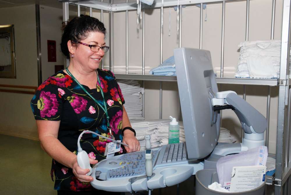
She began her career as an x-ray technologist, eventually moving to her current position as an ultrasound technologist. She’s been in Waterloo Regional Health Network’s medical imaging department since 1989.
How has the medical imaging field changed since you started in the ‘80s?
Where do I start?!
We no longer process images on film which requires a dark room – absolutely everything is digital and goes directly to computers. This allows access to images from virtually anywhere in the hospital.
Also, some tests that used to be done by x-ray are now done by ultrasound instead. For example, we assess leg veins for blood clots using ultrasound imaging, which used to be done by x-ray and required the use of a contract to be injected into the patient’s vein while with ultrasound, there’s no need for an injection and no radiation.
What made you decide to get into this line of work?
My mother used to be a nurse, and suggested that I consider being an x-ray technologist. Back then, CT scans were new and I thought it was really impressive as a modality.
I did end up going to college to be an x-ray technologist, and then eventually an opportunity arose for me to learn ultrasound.
Why are ultrasounds important? What can they detect?
Ultrasounds are important because they can detect pathology without the use of radiation, which can (if used in large amounts) be harmful. Most exams are painless, and can be used on anyone from babies to adults.
An ultrasound is considered “live” imaging as we see everything real-time. We can assess blood flow and its direction, heart motion and even fetal breathing.
They make an image using sound waves instead of x-rays. Ultrasounds detect irregularities like tumors, kidney and gall bladder stones, blocked arteries, blood clots, ligament tears, aneurysms and even abnormalities in fetuses. They can also see the path of a needle, so we help radiologists in doing biopsies as well.
What do you enjoy about your work?
Ultrasounds help diagnose a variety of diseases and abnormalities, and having my own part in that process of identifying irregularities to assist patients is very much rewarding.
Plus, we’re always seeing something new so there’s always something new to learn or examine. The technology constantly advances with new techniques and continually provides more visualized detail.
 Christy Calhoun: treating patients like family
 Wilfred Reck: volunteering to give back
 Marina and Melissa Kaleta: bringing family to the front line of healthcare